Monthly Archives: May 2012

Apert syndrome Part 2

Causes

Acrocephalosyndactyly may be an autosomal dominant disorder. Males and females are affected equally; however research is yet to determine an exact cause. Nonetheless, almost all cases are sporadic, signifying fresh mutations or environmental insult to the genome. The offspring of a parent with Apert syndrome has a 50% chance of inheriting the condition. In 1995, A.O.M. Wilkie published a paper showing evidence that acrocephalosyndactyly is caused by a defect on the fibroblast growth factor receptor 2 gene, on chromosome 10. Continue reading

Pain in the Face and Mouth Part 4

Continued from Part 3

© rozeklaw.com

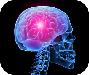
Chronic post-traumatic headache

Most persons who have had head injuries have local pain or tenderness at the site of impact for a few hours or even for a few days, after which many become symptom-free. However up to one half of all persons who injure their heads sufficiently to warrant hospitalization develop chronic post-traumatic headaches. Continue reading

Apert syndrome Part 1

Apert syndrome is a form of acrocephalosyndactyly, a congenital disorder characterized by malformations of the skull, face, hands and feet. It is classified as a branchial arch syndrome, affecting the first branchial (or pharyngeal) arch, the precursor of the maxilla and mandible. Disturbances in the development of the branchial arches in fetal development create lasting and widespread effects. Continue reading

Pain in the Face and Mouth Part 3

Continued from Part 2

© chiro.org

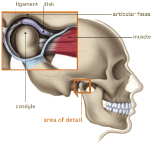
Temporomandibuar joint

Pain from the temporomandibular joint may result from dysfunction, trauma, acute or chronic inflammation, or primary or secondary cancerous tumors. Examination may reveal the masticatory (chewing) muscles tender to palpation or occasionally the joint swollen and warm to touch or tender to palpation via the external auditory meatus. Pain from the temporomandibular joint: Continue reading

Dentinogenesis

Odontoblasts

Dentin-forming cells, odontoblasts, which originate from the ectomesenchyme, form a single layer of cells between the dentin and pulp. The cell body is located on the pulpal wall of dentin and the cellular process extends into the dentinal tubule within the mineralized dentin. The cell bodies are from 3 to 5 m wide and 20 to 40 m long depending on the age of the tooth. The odontoblastic process fills the lumen of the dentin tubule and it is composed of a main trunk, with a diameter of 0.5 to 1 m, and lateral branches. Contrary to the cell body, cell organelles (Golgi apparatus, rough endoblastic reticulum or mitochondria) usually do not appear in the odontoblastic process; however, microtubules, filaments and coated vesicles are present. Odontoblasts are connected to each other with interodontoblastic collagen, the so-called von Korff fibers. Frequent bundles of collagen fibrils enter the odontoblast layer from predentin and are present between odontoblast cell bodies. Ultimately they pass through the odontoblast layer into pulp. Histologically, secretory odontoblasts are columnar in shape. A large number of cytoplasmic organelles are identifiable in young odontoblasts, whereas, aged odontoblasts lose their columnar shape and contain a small number of Golgi apparatus and a small-sized rough endoblasmic reticulum. Continue reading

Leukemia and oral health

What is leukemia?

Leukemia is a malignant cancer of the blood in which one’s bone marrow produces white blood cells in excess in association with gene mutations.

leukemia: white blood cells (dark purple) produced in excess

Causes of leukemia

Genetic predisposition (family history, people with Down syndrome have higher tendency to get leukemia)

Artificial ionizing radiation

Chemicals (eg benzene and other petrochemicals)

Viruses, e.g. HIV (Human Immunodeficiency virus  or HTLV-1 (human T-lymphotropic virus) Continue reading

Pain in the Face and Mouth Part 2

Continued from Part 1

© aafp.org

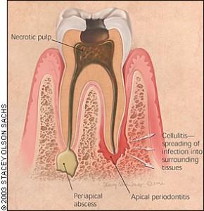
Acute periapical periodontitis

Pain associated with acute perapical periodontitis:

  • Is spontaneous in onset

  • Is moderate to severe in intensity

  • Persists for long periods of time (hours)

  • Is worsen by biting on tooth and in more advanced cases, even by closing the mouth and bringing the affected tooth gently into contact with the opposing teeth. In these cases, the tooth feels ‘high’ (extruded) and is sensitive to touch Continue reading

Pain in the Face and Mouth Part 1

© tna.org.uk

Everyone has experienced pain at least once in their lifetime. Pain is an unpleasant sensory and emotional experience associated with actual or potential damage or described in terms of such damage. Pain is not only a sensory experience, its relation with tissue damage may not be constant and it is often associated with affective and cognitive responses. Continue reading

Amelogenesis

Amelogenesis is the

  1. Amelogenesis is formation of enamel on tooth  and
  2. occurs during the crown stage of tooth development
  3. Amelogenesis occurs after dentinogenesis,

Reciprocal induction.

  1. Since dentin must be present for enamel to be formed,
  2. laying down of dentine induces ameloblasts to secrecte enamel
  3. this is termed reciprocal induction.

Amelogenesis occurs in 3 stages.

  1. The first stage is known as the Pre-secretary  phase,
  2. the second stage is known as the secretary phase and
  3. third stage is called the  maturation stage. Continue reading

Tooth development Part 2

Bell stage

The bell stage is known for the histodifferentiation and morphodifferentiation that takes place. The dental organ is bell-shaped during this stage, and the majority of its cells are called stellate reticulum because of their star-shaped appearance.

THE BELL STAGE IS DIVIDED INTO EARLY BELL STAGE & LATE BELL STAGES. Continue reading