This is a fistulous communication between the floor of the maxillary sinus to the oral cavity. This commonly occurs following dental extraction of infected upper molar and premolar tooth. From a small cavity at birth, the maxillary sinus starts to enlarge during the third month of foetal life and usually reaches maximum development around the eighteenth year. Its volume is approximately 20-25 ml in a normal adult. The floor of the sinus consists of the alveolar process and the hard palate. Continue reading
Monthly Archives: May 2012
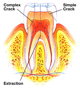
A Cracked Tooth
© clawsonfamilydental.com
Sometimes you feel a sharp tooth pain upon biting and it causes you much annoyance and agony. There are many reasons for pain on biting and a cracked tooth is one of them. Continue reading
Ramsay hunt syndrome
Ramsay Hunt syndrome is a painful rash around the ear that occurs when the varicella zoster virus infects a nerve in the head. This syndrome is also known as geniculate neuralgia or nervus intermedius neuralgia. Ramsay Hunt syndrome can also occur in the absence of a skin rash, condition known as zoster sine herpete. Continue reading
Multiple myeloma
Multiple myeloma (from Greek myelo-, bone marrow), also known as plasma cell myeloma or Kahler’s disease (after Otto Kahler), is cancer of the plasma cells in bone marrow. Continue reading
Kaposi’s sarcoma Part 2
Mouth
Is involved in about 30%, and is the initial site in 15% of AIDS-related KS. In the mouth, the hard palate is most frequently affected, followed by the gums. Lesions in the mouth may be easily damaged by chewing and bleed or suffer secondary infection, and even interfere with eating or speaking. Continue reading
Kaposi’s sarcoma Part 1
Kaposi’s sarcoma (KS) is a tumor caused by Human herpesvirus 8 (HHV8), also known as Kaposi’s sarcoma-associated herpesvirus (KSHV). It was originally described by Moritz Kaposi (KA-po-she), a Hungarian dermatologist practicing at the University of Vienna in 1872. It became more widely known as one of the AIDS defining illnesses in the 1980s. The viral cause for this cancer was discovered in 1994. Although KS is now well-established to be caused by a virus infection, there is widespread lack of awareness of this even among persons at risk for KSHV/HHV-8 infection. Continue reading
Progeria Part 2
A type of anticancer drug, the farnesyltransferase inhibitors (FTIs), has been proposed, but their use has been mostly limited to animal models. A Phase II clinical trial using the FTI lonafarnib began in May 2007. In studies on the cells another anti-cancer drug, rapamycin, caused removal of progerin from the nuclear membrane through autophagy. It has been proved that pravastatin and zoledronate are effective drugs when it comes to the blocking of farnesyl group production. However, it is important to remember that no treatment is able to cure progeria. Continue reading
Progeria Part 1
Progeria (also known as “Hutchinson–Gilford Progeria Syndrome“, “Hutchinson–Gilford syndrome“, and “Progeria syndrome“) is an extremely rare genetic condition wherein symptoms resembling aspects of aging are manifested at an early age. The word progeria comes from the Greek words “pro” (Ï€ÏÏŒ), meaning “before”, and “géras” (γῆÏας), meaning “old age”. The disorder has very low incidences and occurs in an estimated 1 per 8 million live births. Those born with progeria typically live to their mid teens and early twenties. It is a genetic condition that occurs as a new mutation (de novo), and is rarely inherited. Although the term progeria applies strictly speaking to all diseases characterized by premature aging symptoms, and is often used as such, it is often applied specifically in reference to Hutchinson-Gilford Progeria Syndrome (HGPS). Continue reading
Hurler’s Syndrome
Hurler syndrome, also known as mucopolysaccharidosis type I (MPS I), Hurler’s disease, also gargoylism, is a genetic disorder that results in the buildup of glycosaminoglycans (formerly known as mucopolysaccharides) due to a deficiency of alpha-L iduronidase, an enzyme responsible for the degradation of mucopolysaccharides in lysosomes. Without this enzyme, a buildup of heparan sulfate and dermatan sulfate occurs in the body. Symptoms appear during childhood and early death can occur due to organ damage. Continue reading
Chediak-Higashi syndrome
Chédiak–Higashi syndrome is a rare autosomal recessive disorder that arises from a microtubule polymerization defect which leads to a decrease in phagocytosis. The decrease in phagocytosis results in recurrent pyogenic infections, partial albinism and peripheral neuropathy. It occurs in humans, cattle, white tigers, blue Persian cats, Australian blue rats, mice, mink, foxes, and the only known captive albino orca. Continue reading