Apert syndrome is a form of acrocephalosyndactyly, a congenital disorder characterized by malformations of the skull, face, hands and feet. It is classified as a branchial arch syndrome, affecting the first branchial (or pharyngeal) arch, the precursor of the maxilla and mandible. Disturbances in the development of the branchial arches in fetal development create lasting and widespread effects. Continue reading
Tag Archives: mandible
Fracture of the Lower Jaw-Part I
Trauma exerted onto the head and neck region can cause a fracture to any of the bones. The lower jaw, or the mandible, is particularly prone to fracture. In this article we will discuss some of the aspects related to fracture of the lower jaw (mandibular fracture).
CAUSES OF JAW FRACTURE (upper or lower):
- Accidents: Motor-vehicle accidents (MVA), sports injuries, occupational (accidents that occur during work)
- Falls (eg, falling down the stairs, slipping on a slippery floor)
- Assault and fights
- Pathological– a pathology such as tumours or cysts in the jaw bone can cause thinning of the bone or decrease in density of the bone, ultimately leading to fracture of bone in that region, even when a light external force is applied to the bone. Continue reading
Skeletal and dental changes brought by Frankel appliance Part 2
Class II molar relationship
Whether or not there is an increase in size or acceleration of growth of the mandible is one of the major controversies in functional appliance therapy. Although many researchers have claimed that the FR causes extra mandibular growth, this study showed that there were no significant differences between the FR and control groups as far as mandibular movement is concerned, the mean FR movement being 4.1 mm, standard deviation 3.0 mm; the control 5.0 mm, and standard deviation 2.6 mm. As the 5.0-mm change in the control was due to normal growth, it can be assumed that the 4.1-mm change in the FR group was no more than normal growth rather than any effect of the appliance. The maximum value seen in the control was 14.2 mm and for the FR it was 12.8 mm; again, because this change in the controls was due to normal growth it must be assumed that even the maximum FR change was no more than normal growth change. That the size of the mandible is unaffected with the FR is supported by evidence from Creekmore and Radney, and Hamilton et al., who found no significant differences between FR and untreated patient groups. Continue reading
Skeletal and dental changes brought by Frankel appliance Part 1
Functional appliances have been used for many years in the treatment of Class II division 1 malocclusions, the selection of which varies with the type of skeletal and dental anomaly, the growth pattern, and the operator’s preference. Continue reading
Functional appliances
Functional jaw orthpaedics is treatment with functional appliances making use of forces created by the head and neck musculature to bring about the desired dental, facial, and functional changes.
Functional appliances are removable or fixed [cemented] appliances that alter the posture of the mandible [lower jaw] and transmit the forces created by the resulting stretch of the muscles and soft tissues and by the change in the neuromuscular environment to the dental and skeletal tissues to produce movement of the teeth and modification to the growth of the jaws and lower face.
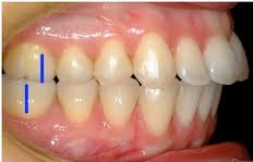
Crossbite
Crossbite is an occlusal irregularity where a tooth (or teeth) has a more buccal or lingual position (that is, the tooth is either closer to the cheek or to the tongue) than its corresponding antagonist tooth in the upper or lower arcade. Continue reading
Muscles of Mastication
During mastication, four muscles of mastication (or musculi masticatorii) are responsible for adduction and lateral motion of the jaw. Other muscles, usually associated with the hyoid such as the sternohyomastoid, are responsible for opening the jaw. Continue reading
Inferior alveolar nerve Part 1
The inferior alveolar nerve (sometimes called the inferior dental nerve) is a branch of the mandibular nerve, which is itself the third branch (V3) of the trigeminal nerve (cranial nerve V). Continue reading
Cherubism Part 2
Anatomy
Cherubism is displayed with genetic conformation and when excessive osteoclasts are found in the affected areas of the mandible and Maxilla. Large cysts will be present with excessive fibrous areas inside the bone. The fibers and cysts will be found among the trabecula of the Coronoid process, the ramus of mandible, the body of mandible and the maxilla regions. The maxilla will be affected up to and including the orbits and sometimes inside the lower orbits. The maxilla and zygomatic bones are depressed and eyes appear to gaze upward. The maxilla has been found to be more severely affected in most cases than the mandible bone. Some patients found with lower inner orbital growths and cysts may lose vision. Continue reading
Cherubism Part 1
Cherubism is a rare genetic disorder that causes prominence in the lower portion in the face. The name is derived from the temporary chubby-cheeked resemblance to putti, often confused with cherubs, in Renaissance paintings.
Presentation
The appearance of people with the disorder is caused by a loss of bone in the mandible which the body replaces with excessive amounts of fibrous tissue. In most cases, the condition fades as the child grows, but in a few even rarer cases the condition continues to deform the affected person’s face. Cherubism also causes premature loss of the primary teeth and uneruption of the permanent teeth. Continue reading