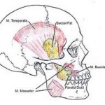
During mastication, four muscles of mastication (or musculi masticatorii) are responsible for adduction and lateral motion of the jaw. Other muscles, usually associated with the hyoid such as the sternohyomastoid, are responsible for opening the jaw.
 Muscles
- The masseter
- The temporalis (the sphenomandibularis is considered a part of the temporalis by some sources, and a distinct muscle by others)
- The medial pterygoid
- The lateral pterygoid
Accessory Muscles Of Mastication:
Suprahyoid Muscles
- Digastric
- Stylohyoid
- Mylohyoid
- Geniohyoid
Infrahyoid muscles
- Sternohyoid
- Thyrohyoid
- Omohyoid
Each of these primary muscles of mastication is paired, with each side of the mandible possessing one of the four.
Now let us discuss in detail about each of these muscles:
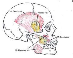
Masseter Muscle:
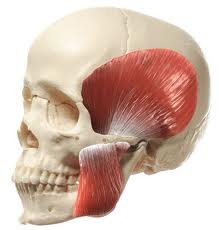
Masseter (below) and temporalis (above) muscle
It is one of the main muscle which helps in the process of mastication.
In humans, the masseter is the second most efficient masticatory muscle. Its origin and insertion make it very useful for the movement of the jaw and for applying good bite force for mastication.
Masseter muscle is a powerful muscle because of its Multipennate arrangement of fibers
The masseter muscle extends from the zygomatic arch to the ramus and body of the mandible. The fibers of this muscle are broad, extending from the region of the second molar on the surface of the mandible to the surface of the ramus.The muscle is divided into 2 parts
- Superficial
- Deep
Origin:
- Superficial layer – anterior 2/3rd of lower border of zygomatic arch & zygomatic processof maxilla
- Middle layer – anterior 2/3rd of deep surface & posterior 1/3rd  of lower border of zygomatic arch
- Deep layer – deep surface of zygomatic arch
Insertion :
- Superficial layer –lower part of lateral surface of ramus of mandible
- Middle layer –middle part of ramus
- Deep layer – upper part of the ramus & coronoid process
The main function of masseter muscle is
- Elevation of the mandible
- lateral movements of the mandible for efficient chewing and grinding of the food
- unilateral  chewing
- Retraction of the mandible
Blood supply:
- Masseteric artery .
Nerve supply:
- Massetric nerve.
Clinical Importance of Masseter Muscle of Mastication:
- Masseter muscle can be palpated both intraorally and extraorally
- Most common muscle involved in Myositis Ossificans
- Masseter Muscle shows Dual action in Complete Denture
- The muscle that commonly undergoes Hypertrophy in Bruxism is Masseter
- Because of the Multipennate arrangement of fibers masseter is a very powerful muscle
Temporalis Muscle:
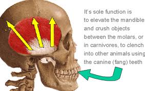 This is the muscle which helps in elevation of the mandible, It is one of the muscles of mastication. It is large shaped in appearance and covers the Temporal area of the skull.
Origin and Insertion:
- From the Parietal bone of the skull and is inserted on the coronoid process of the mandible.
Arterial supply:
- The Deep Temporal artery supplies the large muscle.
Nerve Supply:
- Trigeminal nerve (this nerve has been associated with being the cause of Headache and migrane.)
Functions:
- Elevation of the mandible
- Retraction of the mandible.
- Crushing of food between the molars.
- Posterior fibers draw the mandible backwards after it has been protruded.
- It is also a contributor to side to side grinding movement.
Clinical Importance of Temporalis Muscle:
- Sudden contraction of temporalis muscle will result in coronoid fracture, which is rare.
Lateral Pterygoid Muscle:
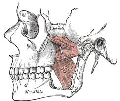
Lateral (horizontal fibers) and medial (vertical fibers) muscle
This is a small muscle which also helps in the mastication process. It is divided into 2 heads
Origin:
- Upper head – infratemporal surface & crest of greater wing of sphenoid bone
- Lower head – lateral pterygoid plate
Insertion :
- Pterygoid fovea on the anterior surface of neck of mandible
- Anterior margin of articular disc & capsule of TMJ
Nerve Supply:
- Pterygoid branch of Trigeminal nerve.
Arterial supply:
- Pterygoid branch of Maxillary artery.
Functions:
- Depresses the mandible
- Protrudes it forward for opening of the jaw
- Side Movements
Clinical Importance of Lateral Pterygoid Muscle:
- Most commonly involved muscle in MPDS
- Only muscle of mastication which has its attachment to the TMJ
- Lateral  Pterygoid forms the roof of the Pterygomandibular space.
Medial Pterygoid muscle:
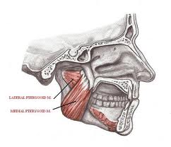 It is a thick muscle of mastication.
Origin and Insertion :
- It  Arises from the deep head the lateral pterygoid plate, and from the maxillary tuberosity.
- Insertion is seen on the Medial angle of the Mandible.
Arterial supply:
- Pterygoid branch of Maxillary artery.
Nerve Supply:
- Mandibular nerve through the medial pterygoid.
Functions:
- Elevates the mandible,
- Closes the jaw,
- Helps in side to side movement.
Clinical Importance of Medial Pterygoid Muscle:
- Medial Pterygoid muscle can be palpated only intraorally
- Most commonly involved in MPDS
- Trismus  following inferior alveolar nerve block is mostly due to involvement of medial pterygoid muscle
Unique features of Masticatory Muscles:
- Have shorter contraction times than most other body muscles
- Incorporate more of muscle spindles to monitor their activity
- Do not have golgi tendon organs to monitor tension
- Elevators predominantly white fibrous which perform fast twitching
- Do not get fatigued easily
- Psychological stress increases the activity of jaw closing muscles
- Occlusal interferences cause a hypertonic synchronous muscle activity
- Closing movement also determined by the height of the teeth