The tongue is composed of muscle tissue with a coating of sensors (dorsal surface) for taste, heat, pain, and tactile information. All the muscles are bilateral, being partially separated by a median septum.
Various muscles of the tongue
Muscles of tongue can be divided into intrinsic and extrinsic groups. Intrinsic muscles are arranged in several planes.
The intrinsic muscles lie entirely within the tongue, while the extrinsic muscles attach the tongue to other structures.
The extrinsic muscles with only terminal fibers inside it, reposition the tongue, while the intrinsic muscles make up most of the mass of the tongue, and alter the shape of the tongue for talking and swallowing.
The front part of the tongue is very flexible and can move around a lot, working with the teeth to create different types of
words. This part also helps you eat by helping to move food around your mouth while you chew. Your tongue pushes the food to your back teeth so the teeth can grind it up.
The muscles in the back of your tongue help you make certain sounds, like the letters “k” and hard “g” (like in the word “go”). Try saying these letters slowly, and you’ll feel how the back of your tongue moves against the top of your mouth to create
the sounds.
The back of your tongue is important for eating as well. Once the food is all ground up and mixed with saliva (say: suh-lye-vuh),
or spit, the back muscles start to work. They move and push a small bit of food along with saliva into your esophagus (say: ih-sah-fuh-gus), which is a food pipe that leads from your throat to your stomach.
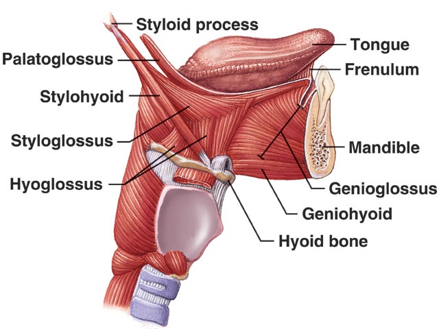
Extrinsic muscles
Extrinsic tongue muscles, by definition, originate from structures outside the tongue and insert into the tongue. The four paired extrinsic muscles protrude, retract, depress, and elevate the tongue:
| Muscle | From | Nerve | Function |
| Genioglossus muscle | mandible | hypoglossal nerve | protrudes the tongue as well as depressing its center. |
| Hyoglossus muscle | hyoid bone | hypoglossal nerve | depresses the tongue. |
| Styloglossus muscle | styloid process | hypoglossal nerve | elevates and retracts the tongue. |
| Palatoglossus muscle | palatine aponeurosis | pharyngeal branch of vagus nerve | depresses the soft palate, moves the palatoglossal fold towards the midline, and elevates the back of the tongue. |

The genioglossus is attached to a point on the mandible about halfway between the chin and the lower incisors (front teeth). (Check by feeling with a forefinger down behind the lower front teeth in your own mouth, and noting where this muscle attaches.) From this point, the muscle fibers fan out posteriorly and superiorly (backwards and upwards) to form the inferior portion of the tongue. The attachment of the genioglossi to the mandible prevents the tongue from falling backward and obstructing respiration.
Anesthetists keep the tongue forward by pulling the mandible forward.
The hyoglossus is a broad, flat muscle running from the greater horn of the hyoid to the posteroinferior lateral aspects of the tongue (on the sides of the lower back of the tongue). It is largely concealed by the mylohyoid muscle. The glossopharyngeal nerve,
stylohyoid ligament, and lingual artery pass deep to the posterior border of the hyoglossus.
The styloglossus extends from the styloid process, a projection of the skull at about the level of the ear down into the lateral aspect of the tongue, interdigitating with the inserting fibers of the hyoglossus.
Palatoglossus muscle is a small muscle that can assist the action of the styloglossus. The palatoglossus muscle can be found on the posterolateral (back, side) aspect of the tongue below the mucous membrane of the anterior faucal pillar (located on the walls
of the mouth on either side of the uvula). From the undersurface of the soft palate, this muscle curves inferiorly (downwards)
to insert into the side of the tongue.
In addition to these four muscles, we will also note two other muscles.
The first is the mylohyoid muscle. It forms the floor of the mouth and controls the raising of the tongue. It is also an elevator of the hyoid bone, and hence elevates the larynx.
The second muscle is the palatopharyngeus muscle, which forms the posterior faucal pillars. It does not affect the position of the tongue. The palatoglossus is innervated by the vagus nerve (the Xth), whereas all the other tongue muscles are innervated by the hypoglossal nerve (the X11th).
Intrinsic muscles
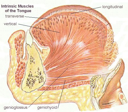 Four paired intrinsic muscles of the tongue originate and insert within the tongue, running along its length. These muscles alter the shape of the tongue by: lengthening and shortening it, curling and uncurling its apex and edges, and flattening and
rounding its surface.
- The superior longitudinal muscle runs along the superior surface of the tongue under the mucous membrane, and elevates, assists in retraction of, or deviates the tip of the tongue. It originates near the epiglottis, the hyoid bone, from the median fibrous septum.
- The inferior longitudinal muscle lines the sides of the tongue, and is joined to the styloglossus muscle.
- The verticalis muscle is located in the middle of the tongue, and joins the superior and inferior longitudinal muscles.
- The transversus muscle divides the tongue at the middle, and is attached to the mucous membranes that run along the sides.