Intense Dry Heat:
In the past method of sterilizing endodontic files was the use of glass bead or salt sterilizer. These sterilizer often need extensive warm-up times and periodic calibration.
Intense Dry Heat:
In the past method of sterilizing endodontic files was the use of glass bead or salt sterilizer. These sterilizer often need extensive warm-up times and periodic calibration.
Definition:
Is defined as freeing the object or substance from all life of any kind
OR
It is defined as the process by which an article, surface or medium is freed of all micro organisms either in the vegetative or spore state Continue reading
CAN BE CLASSIFIED AS:
 1) Distortion
2) Surface roughness and irregularities
3) Porosity
4) Incomplete or missing detail
Our teeth may have different sizes in different individuals, but they all have a typical shape to serve their different functions. Different teeth can be identified from each other by their shapes. However, sometimes abnormalities or variations occur in the shape or form of the tooth, which may be due to developmental causes, or environmental causes such as trauma or genetic causes. Here are some variations in tooth form that one may find in no particular order of occurence:
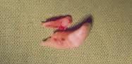
Dilacerated incisor seen from the side view
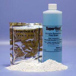
An Investment Material can be described as a ceramic material which is suitable for forming a mold into which metal or alloy is cast. The procedure for forming the mold is described as investing.
Cherubism is a rare genetic disorder that causes prominence in the lower portion in the face. The name is derived from the temporary chubby-cheeked resemblance to putti, often confused with cherubs, in Renaissance paintings.
The appearance of people with the disorder is caused by a loss of bone in the mandible which the body replaces with excessive amounts of fibrous tissue. In most cases, the condition fades as the child grows, but in a few even rarer cases the condition continues to deform the affected person’s face. Cherubism also causes premature loss of the primary teeth and uneruption of the permanent teeth. Continue reading
Macrodontia is derived from the word “macro†which means large and “dont†which refers to tooth or teeth. Macrodontia means a condition whereby a tooth or a group of teeth is abnormally larger than usual. We are not talking about the “bunny teeth” that we had when we were about 6-7 years old, when our 2 upper front teeth has just erupted. The head and jaws of a child that age will continue to grow, and soon the 2 front teeth will no longer seem protruding and large.
Macrodontia can be divided into: Continue reading
Behçet’s disease (Sometimes called Behçet’s syndrome, Morbus Behçet, or Silk Road disease) is a rare immune-mediated systemic vasculitis that often presents with mucous membrane ulceration and ocular involvements. Behçet disease (BD) was named in 1937 after the Turkish dermatologist Hulusi Behçet, who first described the triple-symptom complex of recurrent oral aphthous ulcers, genital ulcers, and uveitis. As a systemic disease, it can also involve visceral organs such as the gastrointestinal tract, pulmonary, musculoskeletal, and neurological systems. This syndrome can be fatal, due to ruptured vascular aneurysms, or severe neurological complications. Continue reading
Oral herpes is an infection of the mouth and lips caused by the herpes simplex virus (also termed HSV). The virus causes painful sores on lips, gums, tongue, roof of the mouth, and inside the cheeks and sometimes on the face and neck. It also can cause symptoms such as fever and muscle aches. People commonly refer to the infection as “cold sores.” Another condition, “canker sore,” is often thought to be caused by HSV, but this is not true. Canker sores occur only inside the mouth, on the tongue and on the soft palate (roof of mouth), not on skin surfaces. Although they reoccur, they are not contagious, usually are self-limiting, and have almost no complications. Canker sores are caused by substances that irritate the oral mucosa. Continue reading
Whenever a lesion is observed on a radiograph, it must first be described in general terms before a differential diagnosis is attempted. Is the lesion radiolucent, radiopaque, or mixed (combination of radiolucency and radiopacity)? Where is the lesion located? The apices of which teeth are involved? What is the size of the lesion? Is the margin of the lesion ill-defined, well-defined, or well-defined with a radiopaque border? Is the appearance of the bone surrounding the lesion: normal, porous, or sclerotic? Continue reading