© elementsofmorphology.nih.gov
Pigmentation in mouth, which ranges from brown to black, may be due to superficial (extrinsic) or deep (intrinsic) causes. They may result from the localization of exogenous substances on or within the mucosa, or may be due to deposition in the tissues of endogenous pigments (for example substances produced by the body), of which melanin is the most common.
Causes of pigmentation around mouth
Exogenous (extrinsic) pigmentation
-
Superficial staining of the mucosa
-
Foreign bodies – amalgam tattoo, miscellaneous
-
Heavy metal salts
Endogenous (intrinsic) pigmentation due to melanin
Developmental causes
-
Racial pigmentation
-
Pigmented nevi
-
Peutz-Jeghers syndrome
Acquired causes
Associated with systemic disease
-
Addison’s disease
-
Pulmonary (lung) disease
-
HIV infection
Associated with hyperkeratosis, chronic inflammation, and lichen planus
-
Drug-induced
-
Idiopathic oral melanotic macule (oral freckle)
-
Lentigo simplex
Neoplastic causes
-
Malignant melanoma in situ
-
Malignant melanoma
Other endogenous pigments
-
Blood breakdown products and disturbances of iron metabolism
Extrinsic staining
Superficial staining of the mucosa
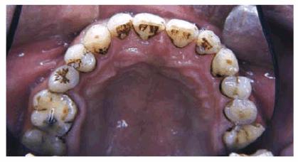
Tobacco stained teeth
Extrinsic discoloration of mouth is rarely of consequence and is usually caused by colored foods, drinks or drugs. Frequently, both mucosa and teeth are discolored. Causes include the following:
-
Foods and beverages, such as beetroot, red wine, coffee and tea
-
Confectionery, such as liquorice
-
Drugs, such as chlorhexidine, iron salts, crack cocaine, minocycline, bismuth subsalicylate and lansoprazole
- Tobacco may cause extrinsic discoloration around mouth, but can also cause intrinsic pigmentary incontinence, this is especially likely in persons who smoke with the lighted end of the cigarette within the mouth (reverse smoking), as practiced mainly in some Asian countries. Tobacco is a risk factor for cancer
- Betel may cause a brownish-red discoloration in mouth, mainly on the teeth and in the cheek mucosa. Betel chewer’s mucosa is not known to be precancerous, but betel use predisposes to submucous fibrosis and to cancer.
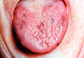
Black hairy tongue
This is a condition in which there is marked increase of thread-like projections on the tongue (filiform papillae), sometimes up to about 1 cm in length, and tongue discoloration associated with overgrowth of pigment-producing bacteria and fungi. The cause is unknown but smoking may be a factor, and in some individuals the disorder follows antibiotic therapy suggesting the disturbance of the normal oral flora is also involved. The condition is usually symptomless but ‘tickling’ of the soft palate may cause gagging and nausea. Black hairy tongue should be distinguished from a simple staining where there is no associated overgrowth of filiform papillae.
Foreign bodies
A variety of foreign substances may be implanted in the oral mucosa giving rise to localized areas of pigmentation. Amalgam is the commonest and best known example but other materials include road grit following road traffic accidents and traumatic implantation of graphite in lead pencil chewers. Some individuals may even have artistic tattooing of the mucosa.
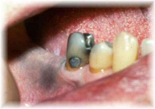
Amalgam tattoo © oralhealthnet.co.uk
Amalgam tattoo
This is a relatively common, often incidental clinical findings, which rarely produces symptoms other than of an area of discoloration. It manifests as a localized blue/black or grayish area and is due to the introduction of amalgam into the soft tissues during such dental procedures at the insertion or removal of restorations, the extraction of teeth when portions of fractured restorations may fall into sockets, and retrograde filling of a root canal after apicectomy. The tattoos may be found in any part of the mouth but are most common in gums or alveolar mucosa of the lower jaw.
Heavy metal salts
Deposits of metallic sulphides in the gum margins are now rarely seen. They may follow absorption of bismuth, lead or mercuric salts either as a result of environmental or occupational exposure or, historically, following therapeutic administration. The salts are present in the fluid found in the gum crevices and are precipitated as sulphides by the action of hydrogen sulphide released as a waste product from plaque organisms. The precipitates cause linear gray or blue/black lines of pigmentation which follow the gum contour around the necks of the teeth. Faint purplish discoloration of the gums may also follow prolonged therapeutic administration of gold compounds, the pigmentation being due mainly to deposition of colloidal metal itself rather than gold salts.
Intrinsic staining
Melanin pigmentation – development causes
Melanin is the commonest of the endogenous pigments in skin and oral mucosa, and is produced by melanocytes present in the basal layer of the epithelium. There is no difference in the number of melanocytes between fair and dark-skinned individuals, the variation in skin and mucosal pigmentation between racial groups being related to differences in activity of melanocytes. The intensity and distribution of racial pigmentation of the oral mucosa is very variable not only between races but also between different individuals of the same race and within different areas of the same mouth. Pigments may be found in any part of the mucosa but the gums is the most common site.
Pigmented nevi are the commonest cause of abnormal melanin pigmentation of skin and every person has a variable number of such lesions, between 20 to 30 on average. These are blue-black often papular lesions formed from increased melanin-containing cells and are usually smaller than 1 cm. However they are relatively rare in oral mucosa and if present they are seen particularly on the palate .
Hypermelanotic pigmentation of skin and/or oral mucosa is also associated with various other development conditions which are not primarily disorders of the melanocyte system.
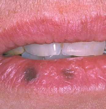
Peutz-Jeghers syndrome lip pigmentation © http://procto.cafe24.com
Peutz-Jeghers syndrome is transmitted through an autosomal dominant gene and is characterized by pigmentation of the mucous membranes and skin, and gastrointestinal polyposis. The polyposis chiefly affects the small intestine and the lesions are not generally considered as pre-cancerous. The melanic pigmentation resembles freckling and appears in infancy. In the mouth the cheek mucosa and lips are usually affected and skin pigmentation occurs rather characteristically around the mouth, nostrils and eyes. Other areas of the skin and other mucosae may be affected. There is a tendency for the skin discoloration to fade in adult life but the mucosal pigmentation persists.
Melanic pigmentation of skin, described as cafe-au-lait spots because of their light brown color, is also seen in some cases of polyostotic fibrous dysplasia and in patients with neurofibromatosis. Pigmentation of the oral mucosa is absent or has only rarely been recorded.
To be continued in Part 2