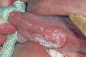
An example of an oral cancer-squamous cell carcinoma
Introduction
Oral or mouth cancers are any tumours that grows anywhere in the mouth. They are often associated with tobacco use. It is a condition of concern because some oral cancers are fatal if not detected and treated early, such as squamous cell carcinoma (SCC), which is due to uncontrolled proliferation of the squamous cells. Almost all oral cancers begin in the flat cells (squamous cell) that cover the surfaces of the mouth, tongue, and lips.
Types
1. Epithelial origin
i) Benign epithelial tumours
-Squamous cell papilloma: usually single, cauliflower-like growths, can be present on any surface of the mouth
ii) HPV induced epithelial hyperplasia
-verruca vulgaris/ common wart: commonly seen in children, can be transmitted from warts on fingers or lips
-veneral wart: usually seen in anogenital areas, but can be found in the oral region if oral sex is practised
-focal epithelial hyperplasia: characterized by multiple small elevated polypoid lesions
iii) Malignant epithelial tumours
-Squamous cell carcinoma: most common of all malignant oral cancers (about 90% of cases)
-Verrucous carcinoma: low grade squamous cell carcinoma (uncommon)
-Basal cell carcinoma: a common tumour on the skin of the face, due to long exposure to UV radiation.
2. Connective tissue origin
Eg: Osteosarcoma, chondrosarcome, rhabdomyosarcoma
3. Odontogenic tumours (tumours formed from tooth-forming tissues)
Eg: ameloblastoma, odontomes
How does tumour form?
Cell growth is regulated by genes:
1. Growth promoting proto oncogenes
2. Growth inhibiting tumour suppressor genes
In health, cellular production and destruction is controlled by a balance between these 2 genes. When there is mutation or deregulation of these genes, this balance is destroyed, resulting in abnormal proliferation of cells or decreased destruction of cells with damaged DNA, forming a tumour.
Â
Predisposing factors/risk factors
1. Tobacco-pipe smokes, cigars, cigarettes, snuff dipping, tobacco chewing, tobacco sachets
2. Betel chewing-betel quid, areca nuts
3. Alcohol-spirits, wines and beers
4. Diet and nutrition-iron deficiency, vitamin A, C, E deficiency
5. Dental factors-sharp teeth, faulty restorations, ill-fiting dentures
6. Ultraviolet light/long term exposure to the sun-on the lip only
7. Viruses-HPV 16 and 18, HSV, HIV
8. Immunosuppression –HIV, organ transplantation
9. Chronic infection-candidiasis, syphilis
10. Occupational

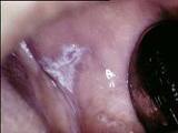
Leukoplakia
Signs and symptoms which may suggest oral cancers
- White patches (leukoplakia) or red patches (erythroplakia) or red &white lesions (erythroleukoplakia) in the mouth
- A mouth sore that won’t heal
- Bleeding in the mouth
- Loose teeth or pathological bony fracture due to underlying bone destruction or invasion
- Problems or pain with swallowing
- A lump in your neck/ lymph node enlargement
- An earache
- Loss of sensation due to infiltration of the tumour into the nerves
Grading and Staging
The purpose of grading and staging is so that there can be a standardized way of classification of tumours between different countries all over the world when communicating the severity of a tumour. It also helps the physician determine the treatment and the prognosis of the patient.
Staging is done clinically, and uses the TNM system:
T-Tumour
T1- Greatest diameter of primary tumour less than or equal 2cm
T2- Greatest diameter of primary tumour more than 2cm but less than 4cm
T3- Greatest diameter of primary tumour more than 4cm
T4- massive tumour more than 4 cm with gross local invasion
N-node
N0- No clinically positive nodes
N1- Single ipsilateral node not greater than 3cm diameter
N2- Single ipsilateral node more than 3cm but less than 6cm
-Multiple ipsilateral nodes less than 6cm
-Bilateral /contralateral nodes less than 3cm
N3- any node more than 6cm
M-metastasis
M0- No distant metastasis
M1- Distant metastasis present
The tumour is staged according to the following criteria:
Stage 1:- T1; N0; M0
Stage 2:- T2; N0; M0
Stage 3:- T3; N0; M0
– T1, T2, or T3; N1; M0
Stage 4:- T4;N0 or N1; M0
– any T; N2 or N3; M0
– any T; any N; M1
Grading
-Grading is done based on the histological features of the tumour (the features of the tumour as seen under a microscope), and can be divided into:
i) Well-differentiated (best prognosis)
ii) Moderately-differentiated
iii) poorly-differentiated (worst prognosis)
Precancerous lesions and conditions
A precancerous lesion is defined as a morphologically altered tissue in which cancer is likely to occur than its normal counterpart. The lesion itself undergoes malignant transformation
Eg:- leukoplakia
-Erythroplakia
-Carcinoma in situ
A precancerous condition is a generalized disorder associated with a significantly increased risk of cancer development some where in the mouth.
Lichen planus is a precancerous condition
Eg: -Oral submucous fibrosis (5-8% chance of occurance of tumour in the mouth)
-Actinic keratosis (10% chance)
-Lichen planus (1-4% chance)
-Conditions associated with epithelial atrophy such as Sideropenic dysphagia
Spread
i) Local invasion
ii) Metastasis to regional lymph nodes
iii) Blood borne metastasis to distant sites