Tag Archives: scaling

Bone Defects and Furcation Involvement Part 2

Continued from Part 1

Diagnosis and treatment planning for bone defects and furcation involvement

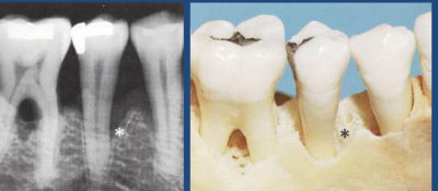
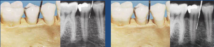
Careful radiographic or x-ray examination is done but it may not reveal the presence of a bone defect or its precise morphology.

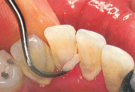
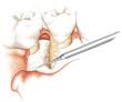
Direct examination of bone morphology:

  • Lift a full thickness mucoperiosteal flap
  • Granulations are curetted and root surfaces planed clean
  • Alveolar crest examined, morphology of bone defect can be defined
  • Mode of treatment decided Continue reading

Bone Defects and Furcation Involvement Part 1

Periodontal disease involves the peridontium consisting of the gums, alveolar or jaw bone, the periodontal ligament and the cementum of tooth. Therefore it is of no surprise if the disease leads to bone defects in our jaw bone or cause furcation involvement. Continue reading

Understanding Dental Instruments ( Part 1 )

Once you enter the dental clinic, you will see a dental chair :

Dental Chair

When you sit down in a dental chair, your eye first goes to the row of shiny, sharp instruments sitting in front of you.  : Continue reading

What to Expect During a Teeth Cleaning Appointment

the cementum is a layer that allows the periodontal ligament attach itself to the root surface

A teeth cleaning appointment is also known as scaling. Every now and then every one of us would need a thorough scaling, because no matter how well we brush and floss and rinse, there will still be some minute debris left in our mouth between our teeth. These debris, after some period of time, will become hardened and these are known as calculus. There are two types of scaling – supragingival scaling, and subgingival scaling. Supragingival scaling is when the dentist only clears away the calculus that are above our gums, and can be seen in plain view. However, subgingival scaling is when the dentists cleans the calculus that are 1 – 2 mm below the gums. Sometimes, scaling is not sufficient for some patients with severe gum conditions, and root planning will be needed. Usually, root planning is needed when there is a gingival pocket more than 4 -5 mm, and there are tenacious calculus and necrotic cementum (refer to the picture to understand what is cementum) stuck to the roots of the teeth. Continue reading

Top 5 Early Signs of Gingivitis

Picture 1: healthy gingiva which is pink, firm, and have knife edges at the neck of the teeth, with mild plaque accumulation

Gingivitis is more commonly known as the inflammation of the gums. The gums are also clinically known as gingiva, hence the term gingivitis. Generally, healthy gums are pink in colour, firm, and do not bleed while brushing. However, it is almost impossible to maintain perfectly healthy gums as gingivitis it is caused by plaque (a thin biofilm of bacteria) that is adhering to our teeth. Plaque can be removed through brushing, but, they are formed again once the brushing stops.  Usually, there is no need to worry about our gum’s health as long as a good brushing regimen is maintained (which is twice daily), however, it does have the potential to evolve into something more destructive, which is known as periodontitis. Continue reading

How to avoid scaling and root planing scam

picture credit to dentalexcellence.co.nz

Scaling? Root planing ? These are everyday dental terms which sort of sound like something used in the field of engineering or construction. To a certain extent, there seems to be a muddle up of understanding about these two terms, what they are, and why they are even needed. This article aims to clear up the air of confusion.

Like nearly all dental problems it all originates from plaque. Plaque is a soft sticky bio-film formed by bacteria which is rather easily cleaned through the use thorough and proper tooth brushing habits. However, should plaque be allowed to build up (due to improper tooth brushing technique or total neglect of oral hygiene.) it may take up trace minerals in ordinary salivary and harden to form what is known as dental calculus, a tenacious solid mass which is nearly impossible to remove through tooth brushing. Without removal of these substances, you are opening the door to gum infection, tooth loss and even serious internal diseases.

Continue reading

A continuation on gingival flap surgery

gingival flap surgery

gingival flap surgery

How is the basic gingival flap surgery procedure performed

First a local anesthetic injection will be given to the site of the surgery to numb the area. This injection is  the same type of injection used when extracting teeth. Therefore, no excessive sedation is necessary unless the patient is very anxious. Then, your dentist will wait a few minutes and test the area again for numbness. The dentist will than make incisions or cuts along the gum margin (neck of the tooth) . When that is done, two vertical incisions are made according to the width  of the gingival flap. The gingival flap is than pulled away from the jaw bone using a forceps and a elevator. Your dentist will then examine the exposed bone and roots.  If there are inflamed or swollen gum tissue between the teeth, your dentist will remove it.  Depending on the situation, inflamed gum tissue may also have to be removed from holes in the bone. Continue reading