Amelogenesis is the
- Amelogenesis is formation of enamel on tooth  and
- occurs during the crown stage of tooth development
- Amelogenesis occurs after dentinogenesis,
Reciprocal induction.
- Since dentin must be present for enamel to be formed,
- laying down of dentine induces ameloblasts to secrecte enamel
- this is termed reciprocal induction.
Amelogenesis occurs in 3 stages.
- The first stage is known as the Pre-secretary phase,
- the second stage is known as the secretary phase and
- third stage is called the  maturation stage.
 In pre-secretary phase,
There is morphogenesis and alignment of secretary cells.
In secretary phase,
- there are histological changes in cells which prepare cells to secret enamel .
- The ameloblasts retreat from the newly formed dentine  and
- laying down of mineralized  enamel with no pre enamel
- The ameloblasts retreat as soon as the enamel is laid down without leaving any process behind
- enamel is laid down in the form of Proteins and an organic matrix to form a partially mineralized enamel in the secretary stage.
In the maturation phase,
- The full thickness of enamel has been laid down
- The excess proteins and water is removed fronm the enamel matrix
- Mineral ions are added   in the enamel matrix
- Growing crystals remove water resulting completes enamel calcification and mineralization.
Stages
Secretory stage
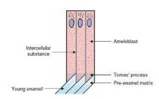 In the secretory stage,
- ameloblasts are polarized columnar cells.
- In the rough endoplasmic reticulum of these cells,
- enamel proteins are released into the surrounding area and
- contribute to what is known as the enamel matrix,
- which is then partially mineralized by the enzyme alkaline phosphatase.
When this first layer is formed,
- the ameloblasts move away from the dentin,
- Allowing for the development of Tomes’ processes at the apical pole of the cell.
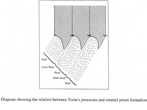
Enamel formation continues,
- around the adjoining ameloblasts,
- resulting in a walled area, or pit,
- that houses a Tomes’ process, and
- also around the end of each Tomes’ process,
- resulting in a deposition of enamel matrix inside of each pit.
The matrix within the pit will eventually become,
- an enamel rod, and
- the walls will eventually become interrod enamel.
The only distinguishing factor between the two is,
the orientation of the calcium crystals.
Maturation stage
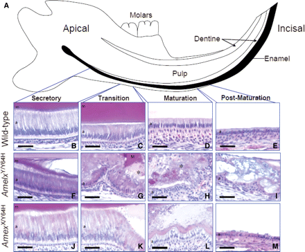
Stages of amelogenesis in a mouse
In the maturation stage,
- the ameloblasts transport substances used in the formation of enamel.
- Microscopically, the most notable aspect of this phase is that these cells become striated, or have a ruffled border.
These signs demonstrate that the ameloblasts have changed their function from,
Production, as in the secretary stage, to transportation.
The noteworthy proteins involved are ,
- amelogenins,
- ameloblastins,
- enamelins, and
- tuftelins.
During this process,
- amelogenins and
- ameloblastins are removed after use,
- leaving enamelins and tuftelin in the enamel.
- By the end of this stage, the enamel has completed its mineralization.
Neo natal lines in enamel:
1.  Formed as a result of trauma of birth
2.  Are excentuated incremental lines
3.  Formed as the anamel rods the prism direction and thickness is changed
Prisms do not follow a straight path from dentino enamel junction to the surface.
1.  Enamel prisms are perpendicular to dentine enamel junction to the surface
2.  Their direction reflect the direction of movement of ameloblast during enamel formation
Hunter –Scherger bands
Their changing direction and arrangement makes,
Hunter –Scherger bands
Changing direction of enamel prism between different layers makes enamel:
1.  Increases strength of enamel
2.  Less prone to fracture
3.  More resistant to wear
Dentino enamel junction:
1.  Is not a straight line but is interlocking of 2 tissues
2.  This prevents shearing apart of the 2 tissues during mastication
3.  The dentino enamel junction appearnce is Scalloped
4.  Where the concavities of scalloped project in to the dentine
This scalloped appearance is prominent:
1.  Beneath the cusps
2.  Enamel and dentine crystals difference
3.  Hydroxyappatite crystals in dentine are smaller then enamel
Histological structures apper at the dentino enamel junction:
- Enamel spindles
- Enamel tufts
- Enamel lamella
Enamel tufts
Enamel tufts:
- Are found in the inner 1/3 of enamel
- Attached to enamel dentine junction
- Are hypo mineralised
- Recur at intervals
- Have more organic matrix
Enamel lamellae:
- Are hypomineralised sheets
- Run full length of enamel
- Are less common then tufts
- Are irregularly arranged
Enamel spindles:
- Are the projection of dentinal tubules which cross dentino enamel junction and pass in to denamel
- And appear as enamel spindles
- They project 10 – 40 micron meter in to enamel
- They are common beneath cusps and sdges
- The spindles sre continuous with the dentinal tubules
Normally the dentinal tubules terminate at the:
- Dentine enamel junction
- But some of the dentinal tubules enter the enamel and appear pore like structures microscopically  appear as enamel spindles
Dentinal tubule:
Contain odontoblast process
Surface enamel:
- Is physically and chemically different from the sub- surface enamel
- Surface enamel is harder
- Less porous
- Less soluble
- More radio- opaque then sub surface enamel
Surface enamel has a wavy surface:
- Called perikymata
- On newly erypted tooth
- With elevations called imbrications and
- Furrows prikymata