Tooth development or odontogenesis is the complex process by which teeth form from embryonic cells, grow, and erupt into the mouth. Although many diverse species have teeth, non-human tooth development is largely the same as in humans. For human teeth to have a healthy oral environment, enamel, dentin, cementum, and the periodontium must all develop during appropriate stages of fetal development. Primary (baby) teeth start to form between the sixth and eighth weeks, and permanent teeth begin to form in the twentieth week. If teeth do not start to develop at or near these times, they will not develop at all.
Credits to: Nature revies @ Genetics
A significant amount of research has focused on determining the processes that initiate tooth development. It is widely accepted that there is a factor within the tissues of the first branchial arch that is necessary for the development of teeth.
In vertebrates several specializations of epithelial tissue (‘phanères’) generate after thickening specific structures: keratinized structure (hair, nails) or exoskeletons structure (scales, teeth). Placoids scales and teeth of sharks are considered homologous organs.
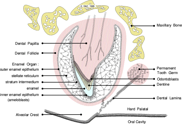
The tooth bud (sometimes called the tooth germ) is an aggregation of cells that eventually forms a tooth. These cells are derived from the ectoderm of the first branchial arch and the ectomesenchyme of the neural crest. The tooth bud is organized into three parts: the enamel organ, the dental papilla and the dental follicle.
The enamel organ is composed of the outer enamel epithelium, inner enamel epithelium, stellate reticulum and stratum intermedium. These cells give rise to ameloblasts, which produce enamel and the reduced enamel epithelium. The location where the outer enamel epithelium and inner enamel epithelium join is called the cervical loop. The growth of cervical loop cells into the deeper tissues forms Hertwig’s Epithelial Root Sheath, which determines the root shape of the tooth.
 The dental papilla contains cells that develop into odontoblasts, which are dentin-forming cells. Additionally, the junction between the dental papilla and inner enamel epithelium determines the crown shape of a tooth. Mesenchymal cells within the dental papilla are responsible for formation of tooth pulp.
The dental follicle gives rise to three important entities: cementoblasts, osteoblasts, and fibroblasts. Cementoblasts form the cementum of a tooth. Osteoblasts give rise to the alveolar bone around the roots of teeth. Fibroblasts develop the periodontal ligaments which connect teeth to the alveolar bone through cementum.
The developing tooth bud
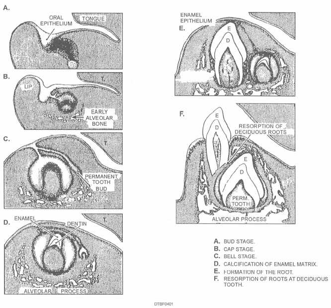
One of the earliest steps in the formation of a tooth that can be seen microscopically is the distinction between the vestibular lamina and the dental lamina. The dental lamina connects the developing tooth bud to the epithelial layer of the mouth for a significant time.
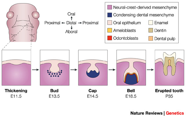
Tooth development is commonly divided into the following stages: the bud stage, the cap, the bell, and finally maturation. The staging of tooth development is an attempt to categorize changes that take place along a continuum; frequently it is difficult to decide what stage should be assigned to a particular developing tooth. This determination is further complicated by the varying appearance of different histologic sections of the same developing tooth, which can appear to be different stages.
Bud stage
Credits to: Dacross services
The bud stage is characterized by the appearance of a tooth bud without a clear arrangement of cells. The stage technically begins once epithelial cells proliferate into the ectomesenchyme of the jaw. Typically, this occurs when the fetus is around 6 weeks old. The tooth bud itself is the group of cells at the end of the dental lamina.
Along with the formation of the dental lamina, 10 round epithelial structures, each referred to as a bud, develop at the distal aspect of the dental lamina of each arch. These correspond to the 10 deciduous teeth of each dental arch, and they signify the bud stage of tooth development. Each bud is separated from the ectomesenchyme by a basement membrane. Ectomesenchymal cells congregate deep to the bud, forming a cluster of cells, which is the initiation of the condensation of the ectomesenchyme. The remaining ectomesenchymal cells are arranged in a more or less haphazardly uniform fashion.
Cap stage
The first signs of an arrangement of cells in the tooth bud occur in the cap stage. A small group of ectomesenchymal cells stops producing extracellular substances, which results in an aggregation of these cells called the dental papilla. At this point, the tooth bud grows around the ectomesenchymal aggregation, taking on the appearance of a cap, and becomes the enamel (or dental) organ. A condensation of ectomesenchymal cells called the dental follicle surrounds the enamel organ and limits the dental papilla. Eventually, the enamel organ will produce enamel, the dental papilla will produce dentin and pulp, and the dental follicle will produce all the supporting structures of a tooth.