PRINCIPLES IN CHOOSING A FILTER MATERIAL
The material chosen must attenuate principally by means of photoelectric effect in the photon energy range being dealt with in order to discriminate against low energy photons
The material must not have an absorption edge close to the energies of the photons that it is desired to use
The thickness of the material must not be too small – it should be uniform
Too thin might produce pin holes which might produce completely unfiltered beams
Filtration affects beam quality/energy/penetrating ability
a)Â Â Â Â Â Â Average beam energy/penetrability
i)Â Â Â Â Â Â Â Â Depends on:
(1)Â Â Â Â kVp
(2)Â Â Â Â Amount of total filtration in the beam
ii)Â Â Â Â Â Â kVp also determines the minimum wavelength of the beam
iii)Â Â Â Â Â Filtration determines the maximum wavelength of the beam
iv)Â Â Â Â Increasing either kVp or filtration will increase the average energy of the beam, allowing it to be more penetrating and of higher quality
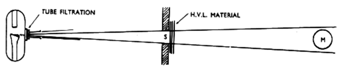
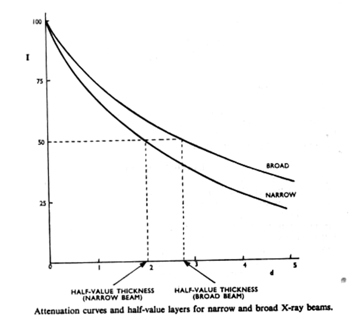
b)Â Â Â Â Â Â Beam quality is measured by its half-value layer (HVL)
HVL: that thickness of a specified material (usually a metal) which reduces the exposure rate to one-half its initial value
It is the most common method to measure radiation quality i.e. penetrating power
Federal regulation states that at 80 kVp, the half-value layer must be 2.34 mm equivalent
HVL: that thickness of a specified material (usually a metal) which reduces the exposure rate to one-half its initial value
It is the most common method to measure radiation quality i.e. penetrating power
Federal regulation states that at 80 kVp, the half-value layer must be 2.34 mm equivalent

The filter materials and H.V.L. materials used are generally the same , the difference being that the filter is a permanent feature of the tube whereas the H.V.L. material is part of the measuring apparatus
c)Â Â Â Â Â Â Because filtration causes the beam to be more penetrating, increasing filtration:
i)Â Â Â Â Â Â Â Â Decreases density
ii)Â Â Â Â Â Â Decreases exposure rate
iii)Â Â Â Â Â Decreases contrast
TYPES
INHERENT FILTERS
ADDED FILTERS
TOTAL FILTERS
INHERENT FILTERS
ADDED FILTERS compensation and
equalization filters.
TOTAL FILTERS
Examples of compensation filters are variations of the wedge filter, which is used to compensate for the otherwise uneven X-ray fluence generated by objects with a wide thickness variation, such as hands and feet.
Equalization filters follow a similar principle and are sometimes used to compensate for more irregular absorption variations in the object, such as the mediastinum in a chest frontal image.
Inherent filtration
the filtration of an X-ray beam by any parts of the X ray tube or tube shield through which the beam must pass.
The parts include
the glass envelope of the X-ray tube,
the oil cooling the tube and
the exit window in the tube housing.
The inherent filtration corresponds to approximately 0.5–1 mm of aluminium.
The total filtration of the X-ray beam before it reaches the patient consists of the inherent filtration plus the added filtration.
ADDED FILTERS
It is the external filtration supplied in the form of discs placed over the port in the head of the x-ray machine
The purpose is to filter out longer wavelength low energy x- rays from the beams
commonly metallic filters inserted into the X-ray beam.
made of either aluminium or copper
Total filtration
It is the sum of the inherent filtration plus any added external filtration supplied in the form of Al discs placed over the port in the head of the x- ray machine
Federal regulation requires the total filtration to be –
1.5mm of Al for 70 kvp
2.5mm of Al for higher voltage
Compensators
Compensators are added filtration with a shape intended to change the spatial pattern of the x-ray intensity incident on the patient, so as to deliver a more uniform x-ray exposure to the detector
Placed close to the x-ray tube port or just external to the collimator assembly
May be made of metal or a plastic compound (EX: boomerang)
ii)Â Â Â Â Â Â Used where there is difficulty imaging body parts due to varying tissue thickness and composition
ALUMINIUM
LEAD
TIN
COPPER
LOW ENERGY BEAMS
Used in diagnostic radiology
It is generated at 80 kv
Points to be noted-
quite a lot of radiation has an energy of 20 kev or less so it does not penetrate through the patient to play a part in the production of radiograph .
These low energy photons should therefore be removed
ALUMINIUM (Z=13)
Beams used for diagnostic purposes are in the range of 25- 50kev.
K level of Al is 1.6kev (well below the lowest photon energy in this spectrum), so is most suitable for filtration of this range.
Much of the attenuation is by compton effect, rather than photo electric effect.
It is even less effective than Cu