© southridingfamilydentistry.com
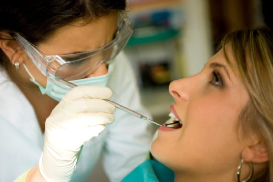
Sometimes you may hear the term ‘biopsy’ when investigations are required for certain lesions in the mouth. Biopsy sounds like a scary procedure that brings the word ‘cancer’ to mind but in fact it is just an aid for diagnostic purposes.
What is biopsy?
Biopsy is the removal of tissues from a living individual for diagnostic examination.
What are the indications for biopsy?
- Any lesions or ulcer that persists for more than 2 weeks with no apparent cause
- Any inflammatory lesion that does not respond to local treatment after 10 to 14 days (after removing the irritant)
- Persistent hyperkeratosis (overgrowth of layers of skin) changes in surface tissues
- Any persistence lesion, either visible or palpable beneath relatively normal tissue
- Inflammatory changes of unknown cause that persist for long periods
- Lesions that interfere with local function (for example fibroma)
- Bone lesions not specifically identified by clinical and radiographic findings
- Any lesion that has the characteristics of malignancy
Types of biopsy
- Oral cytology – exfoliative cytology, oral brush biopsy
- Aspiration biopsy – fine needle aspiration cytology
- Incisional biopsy
- Excisional biopsy
Exfoliative cytology
It is a diagnostic tool for tumor cells (used generally for uterine cervical malignancy). Although application to oral cavity has been advocated, it should be used as an adjunct to, not a substitute for incisional or excisional biopsy.
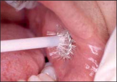
Oral brush biopsy
© internalmedicinenews.com
Indications
- Indicated for chronic mucosal changes such as leukoplakia, lichen planus and patients with a history of oral cancer
- chair side test,
- do not require any topical or local anesthesia,
- minimal discomfort and bleeding,
- only seconds to perform
Advantages
Procedure
- A special brush to collect the epithelial cells.
- The brush is placed in contact with oral epithelium and rotated with firm pressure 5 to 10 times.
- Properly performed, the brush collects cells from all three layers of the epithelium – basal, intermediate, superficial layers.
- The cellular material collected on the brush is transferred to a glass slide and flooded with fixative.
- After the slide is dry, it is sent to a special lab where the pathologist will determine whether the brush has penetrated the basement membrane.
- If the biopsy has not collected cells from the full thickness of epithelium, biopsy has to be repeated.
Oral cytology is not a substitute for traditional scalpel biopsy. Oral lesions with interpretations as positive or atypical require scalpel biopsy to characterize the lesion completely.
Aspiration biopsy

© scielo.br
Aspiration biopsy is the use of a needle and syringe to penetrate a lesion in order to aspirate its content to determine whether air or fluid is present.
Fine needle aspiration cytology is a procedure whereby the pathologist uses a special needle and syringe to enter the tissue and collect cells for histological examination. Commonly used in salivary gland tumors.
Indications
Aspiration should be carried out an all lesions thought to contain fluid (with the possible exception of a mucocele) before a surgical exploration.
Technique
- An 18 gauge biopsy needle is connected to a 5 to 10 ml syringe
- The area is anesthetized and the 18 gauge needle inserted into the depth of mass during aspiration
- The tip of needle may have to be repeatedly repositioned in an effort to locate a fluid center
- For lesion within bone biopsy, if expansion and thinning of the jaw bone has occurred, the needle may be firmly applied directly to bone and twisted until it perforates the cortical plate of the jaw bone
Incisional biopsy
Incisional biopsy is removal of a part of the entire pathological tissue at the time of surgery.
Indications
- Lesion more than 1 centimeters in diameter
- Lesions at hazardous location
Excisional biopsy
Excisional biopsy is removal of the entire pathological tissue at the time of surgery.
Indications
- Should be used with smaller lesions (less the 1 centimeters in diameter)
- Any lesion that can be removed entirely without mutilating the patient
- Pigmented and small vesicular lesions
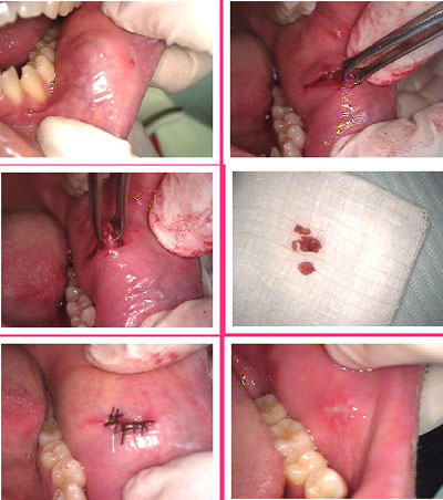
Surgical biopsy procedure
© nycdentist.com
- Your informed consent is needed before the biopsy can take place.
- Local anesthesia solution will be injected in the area of concern.
- The soft tissues surrounding the area of concern are stabilized.
- A sharp scalpel is used for incision of the lesion.
- Two incisions forming an ellipse at the surface and converging to form a V at the base of the lesion provide a good specimen and leave a wound that is easy to close.
- A periphery of normal appearing tissues is included in biopsy specimen. For benign lesions 2 to 3 millimeters of peripheral tissues is required. For malignant, vascular, or pigmented lesion 5 millimeters of peripheral tissue is needed.
- After removal of the tissue it is placed immediately in 10% formalin solution that is at least 20 times the volume of the surgical specimen.
- Primary surgical closure is indicated at the wound area. The mucosa is undermined with the help of pointed scissors. This allows the approximation of tissues with little tension on the suture line.
- The dentist will inform you of the biopsy results which should be obtained within a week or two.