Continued from Part 1
© dentistryandmedicine.blogspot.com
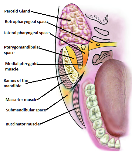
- Abscesses in the molar region of either jaw may penetrate the buccal cortical plate above (in the upper jaw) or below (in the lower jaw) the attachments of the buccinators muscle (A muscle that flattens the cheek and retracts the angle of the mouth). In such acute inflammatory edema and pus discharge spread into the soft tissues of the face or neck. This may present as a cellulitis or less frequently as a localized soft tissue abscess depending on the nature of the infection. Such an abscess may track towards the overlying skin to discharge through a sinus on the skin surface. The abscess may then become chronic with the sinus discharging pus periodically, associated with increasing fibrosis, scarring and disfigurement.
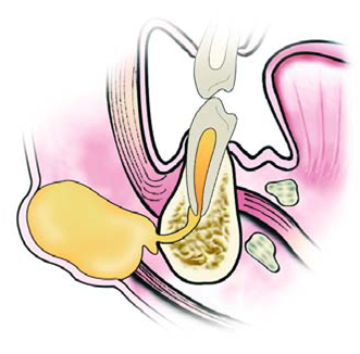
- An abscess related to a lower premolar or molar tooth may perforate the lingual plate of the lower jaw below the attachment of the mylohyoid muscle to involve the submandibular space. This causes a marked swelling at the lower border of the lower jaw spreading towards the neck. The submandibular space has communications with the sublingual and lateral pharyngeal space, into which the infection may subsequently spread. An abscess in the submandibular region is separated from the skin by the deep fascia of the neck and so tends to track in a front and back direction under the skin surface.
Subcutaneous abscess © dentistryandmedicine.blogspot.com
- Pus from an abscess associated with a lower incisor or canine may track labially and perforate the bone below the insertion of the mentalis muscle and pass downwards to present as a subcutaneous abscess, most often in the midline between the points of attachment of the two mentalis muscles.
What is the difference between abscess and cellulitis?
An abscess is a localized collection of pus which feels hard if small, tense or covered by a thick layer of tissues. If large it may be softer and exhibit fluctuance (movement or sway in a rising and falling or wavelike pattern). Pointing to the skin or mucosa indicates mouth abscess formation.
Cellulitis is a rapidly spreading inflammation of the soft tissues particularly associated with streptococcal infections. It is not well-localized, brawny swelling in contrast to a circumscribed abscess in mouth, and the rapid spread is most likely related to the release of large amounts of streptokinase and hyaluronidase which are produced by most strains of streptococci.

© toothfairy2.com
Clinically, there is diffuse, tense, painful swelling of the involved soft tissues usually associated with physical discomfort and an elevated temperature. Much of the swelling is due to inflammatory edema; pus discharge and abscess formation occur later if treatment is neglected or delayed. Cellulitis associated with upper teeth initially involves the upper half of the face. Extension towards the eye is a potentially serious complication because of the risk of cavernous sinus thrombosis as a result of infection involving veins at the inner canthus of the eye which communicate with the cavernous sinus.
Cellulitits associated with lower teeth initially involves the lower half of the face; extension into the submandibular and cervical tissues may cause respiratory embarrassment. Cellulitis spreading into the deeper surgical spaces usually presents clinically as pain and difficulty in opening the mouth rather than facial swelling.
What is cavernous sinus thrombosis?

© dentallecnotes.blogspot.com
Thrombosis (formation or presence of a clot of coagulated blood attached at the site of its formation in a blood vessel) of the cavernous sinus is a serious life threatening condition. It follows spread of odontogenic infection along two main venous pathways. Bacteria and the infected clot travel towards the back from the upper lip and face via the anterior facial vein. Alternatively infection may spread from the pterygoid space via the pterygoid plexus of veins. In addition to treatment the for infection, thrombosis requires anticoagulation. The mortality rate is high.
Local features are seen on one side at first but the signs become both sides as the thrombus or clot grows. Local effects include marked swelling of the eyelids, pulsating protrusion of the eyeball from the socket, a dilated facial vein, restriction of movement of the eyeball, and bleeding in the retina. Systemic effects include rapid pulse, marked fever and severe physical discomfort.
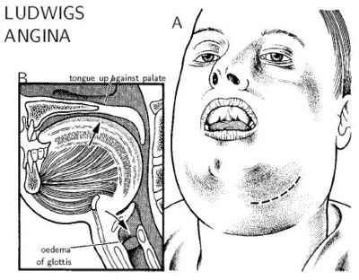
What is Ludwig’s angina?
© ps.cnis.ca
Ludwig’s angina is severe cellulitis involving the submandibular, sublingual and submental space on both sides of the face, usually as a result of initial involvement of the submandibular space. Since the advent of antibiotics it is now rare.
The diffuse cellulitis produces a board-like swelling of the floor of the mouth, the tongue being elevated and displaced towards the back. As a result there is difficulty in opening the mouth, eating, swallowing and breathing. The latter is exacerbated as infection tracks backwards to involve the pharynx and larynx. Edema of the glottis may occur with risk of death by suffocation. This condition requires immediate management, drainage of abscess and aggressive antibiotic therapy.