Impacted Maxillary Canines
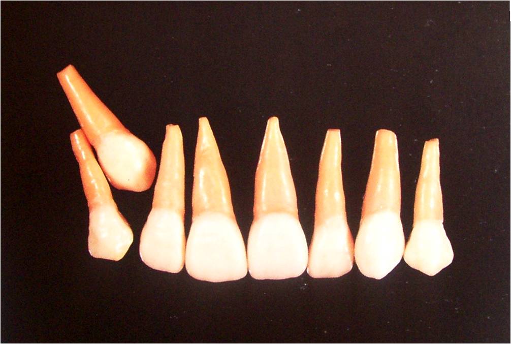
Natural Development of Canine
Why do maxillary canines are frequently impacted?
Etiologic factors
A summary of Dewel’s discussion of these factors follows –
The hard palatal bone offers more resistance than does alveolar bone on the ridge to the eruption of the cuspid that is misplaced lingually.
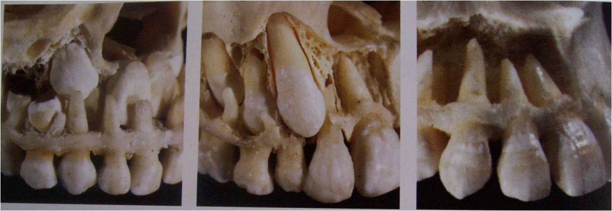
The greater the distance the tooth must travel from its point of development to normal occlusion, the greater the possibility of deflection from its normal course and of its resultant impaction.
During development the crown of the permanent cuspid lies immediately lingual to the long apex of the primary cuspid root. Any change in position causes impaction.
Delayed resorption of the primary cuspid root.
Cuspids erupt between teeth already in occlusion and are competing for space with the also currently erupting second molar.
Cuspid is preceded by a primary cuspid whose mesiodistal diameter is much less than that of the permanent cuspid.
Clinical Examination for Canine Impaction
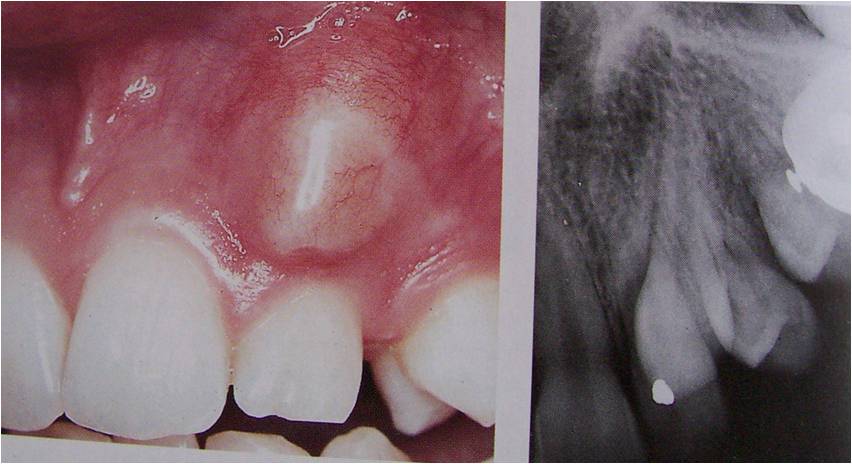
“Clinical palpation of the buccal surface of the alveolar process distal to the lateral incisor may reveal the position of the maxillary canine about 1 to 1½ years before emergence and this has been suggested as a diagnostic toolâ€.
Clinical Diagnosis
Signs of canine eruption
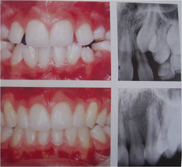
Upper : Distal tipping of a lateral incisor due to the erupting canine
Lower : Follow-up – Straightening of the teeth after canine eruption
Radiographic diagnosis
When should we suspect canine impaction?
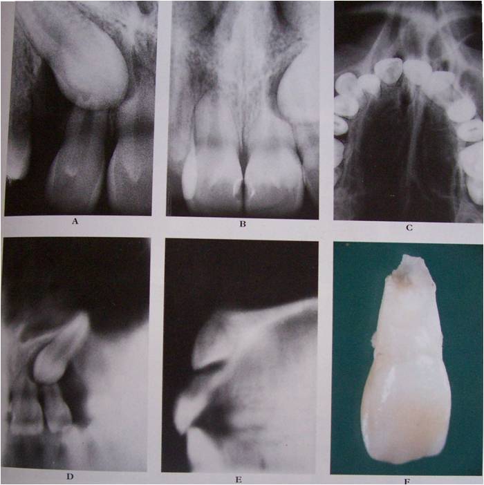
Critical period – 8 to 11 years
Radiographic examination mandatory if there is no eruption after the critical age.
11 years appears to be the critical age.
Treatment Options for Impacted Canine
No treatment of the unerupted canine
Prophylactic space augmentation
Extraction of primary canine (Guided eruption)
Surgical repositioning
Removal of the canine
Surgical Exposure & Bracket Placement for guided eruption
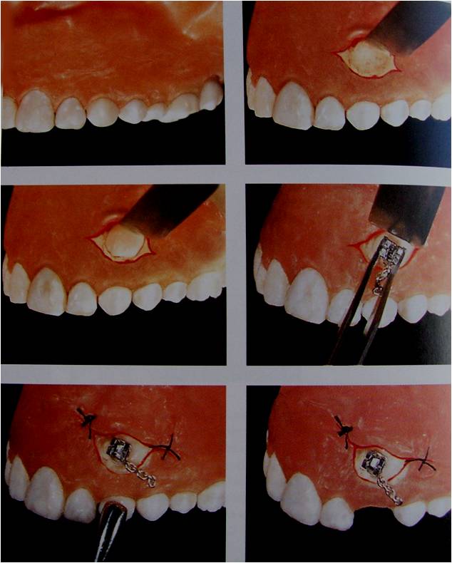
Surgical uncovering canine and exposure
Removal of bone and follicle
Extraction of primary canine and bracket placement
Surgical Removal of Palatally Impacted Canine
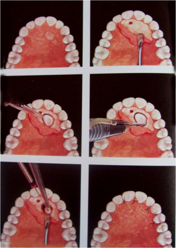
Exposure of the palatally positioned canine via paramarginal incision and fenestration
Surgical uncovering of the crown
Splitting of the crown and separation of the root
Repositioning the flap and suturing
Indications for Canine Transplantation
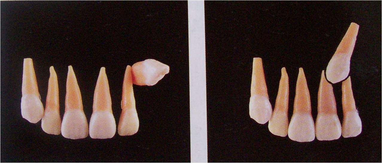
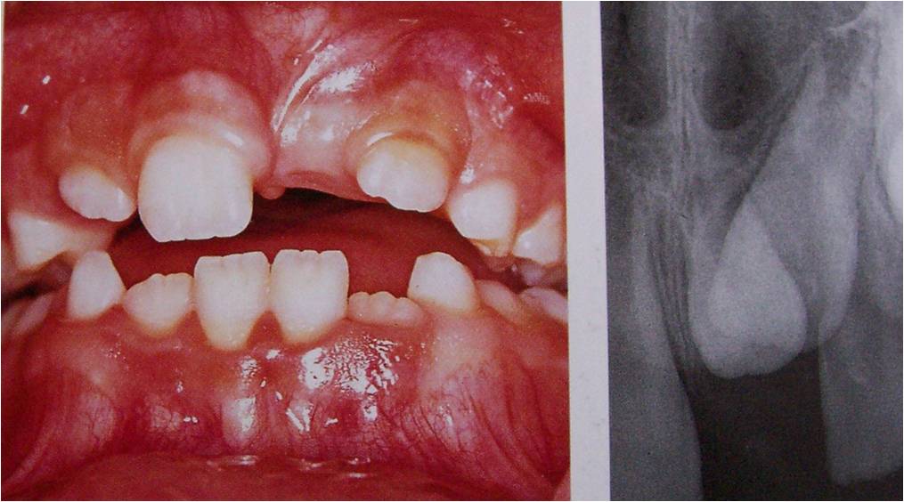
Left : High labial ectopic position
Right : Close proximity to the apices of the adjacent teeth.
Transplantation of Labially Positioned Canine
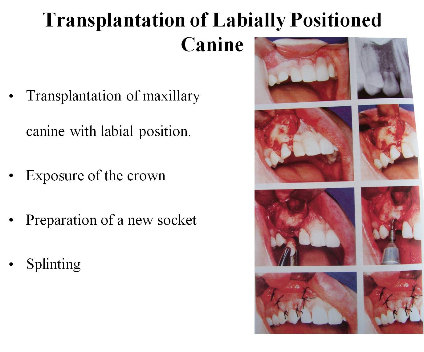
Transplantation of maxillary canine with labial position.
Exposure of the crown
Preparation of a new socket
Splinting
Supernumerary Tooth
It is an extra tooth that appears in the mouth other than the normal number of teeth that is supposed to be in the mouth
What will happen if we leave a supernumerary tooth?
Either eruption / impaction?
Periodontitis
Spontaneous resorption (freq 8%)
Migration
Follicular/dentigerous cyst (2% to 9%)
Warnings for disturbance in eruption of anterior permanent teeth
In the central incisor region a first indication of disturbance in eruption is an altered timing of eruption of the central incisor.
The next warning is the eruption of lateral incisors ahead of the centrals.
Timing of supernumerary tooth removal
Removal before the age of five years resulted in significantly fewer eruption problems of permanent incisiors compared to removal at 7 years of age or later
The Impacted Tooth in Patients with Alveolar Clefts
The Impacted Tooth in Patients with Alveolar Clefts
Tooth impaction is a relatively common problem in individual with clefts of the lip and palate.
Impactions are not confined to the site of cleft.
Canine impactions to be 10 times more frequent in clefts than in non cleft individuals.
Striking anomalies of dental development are found in the maxillary lateral incisor region where the alveolar cleft occurs.
Lateral incisor being the most affected.
Cleft and Canine
When cleft subtype was taken into consideration, it was apparent that the great majority of patients with impacted canine were those with unilateral cleft lip and palate (UCLP)
Impaction frequency is much lower in bilateral cleft lip and palate (BCLP).
Alveolar Bone Grafting and Tooth Impaction
It has been suggested that grafts of cancellous bone may delay / impede canine eruption. Incidence – 3 to 73%
Causes for increased frequency of canine impaction in bone grafting.
Bone graft may actually change of path eruption of cleft side canine
Mucobuccal flaps used to cover alveolar margin, the tissues abundant elastic fibers and loose connective tissue represent an obstacle to tooth eruption.
Surgical Considerations for impacted tooth in the region of elective osteotomiesThere seems to be a dichotomy of opinion whether to remove impacted teeth prior to the orthognathic surgery or at the actual time of surgery.
The following points address this issue and systematically outlines practical and biologically sound methods of managing impacted teeth in the path of planned osteotomies.