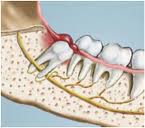
Pericoronitis is  a dental disorder in which gum tissue around molar teeth/last teeth/wisdom teeth becomes swollen and infected. It is an acute infection which refers to inflammation of gingiva and surrounding soft tissues of an incompletely erupted tooth. It occurs most frequently in the mandibular third molar area.
Acute inflammation is a short-term process, usually appearing within a few minutes or hours and ceasing upon the removal of the injurious stimulus. It is characterized by five cardinal signs.
The acronym that may be used for this is “PRISH” for Pain, Redness, Immobility (loss of function), Swelling and Heat.
The traditional names for signs of inflammation come from Latin:
Dolor means Pain
Calor means Heat
Rubor means Redness
Tumor means Swelling
Functio laesa means Loss of Functions
Clinical Features :
Marked red,edematous suppurating lesion that is extremely tender with radiating pain to the ear, throat and floor of the mouth
Extremely uncomfortable because of the foul taste and inability to close the jaws
Swelling of the cheek in the region of the angle of the jaw
Acute Pericoronitis
It is identified by varying degrees of involvement of pericoronal flap as well as with systemic complications. An influx of inflammatory fluid and cellular exudates results in an increase in bulk of the flap which interferes with complete closure of the jaws. The flap is traumatized by contact with the opposing jaw and inflammatory involvement is aggravated.
Lymphadenitis is common. Toxic systemic complications such as fever, leukocytosis and malaise are present.
Complications
The involvement may become localized, in the form of pericoronal abscess.
If it  occurs in a partly-erupted vital tooth , it may give rise to cyst formation.
It may spread posteriorly into the oropharyngeal area and medially into the base of the tongue, making it difficult for the patient to swallow.
Depending on the severity, there is involvement of the submaxillary, cervical, deep cervical and retropharyngeal lymph node. Peritonsillar abscess formation, cellulitis and Ludwig’s angina are infrequent but nevertheless potential sequelae of acute pericoronitis.
Treatment
The treatment of pericoronitis depends on:
- Severity of the inflammation
- The systemic complications
- The advisability of, retaining the involved tooth
Pericoronitis can be tricky to treat. That’s because the flap of gum tissue won’t go away until the wisdom tooth emerges naturally, the tissue is removed or the tooth is removed. Your dentist will clean the area thoroughly by rinsing under the flap with water to remove bits of food and pus. Your dentist also may need to remove damaged tissue. If the area is infected, you’ll most likely be given antibiotics.
Your dentist will explain how to keep the area clean, which is the best way to prevent the problem from returning. This usually involves brushing and flossing daily and rinsing your mouth with water several times a day. These steps will help to prevent food from getting stuck under the gum flap.
In some cases, your dentist may suggest removing the erupting tooth. Or the dentist may want to remove the tooth above it, which bites down on the gum below. If your dentist thinks the tooth may erupt fully into the mouth without problems, he or she may leave it alone. However, if pericoronitis comes back, the tooth may be extracted.
Pericoronitis that causes symptoms should be treated as soon as possible. If it is not, the infection can spread to other areas of your mouth. The most severe cases are treated in a hospital. They sometimes require intravenous antibiotics and surgery.
First Visit
- The area is gently flushed with warm water to remove superficial debris and exudate followed by application of topical anesthetic agent
- The flap is reflected with a scaler and the underlying debris is also removed and the area is flushed with warm water
- Â Instructions to the patient include hourly rinses with a solution of a tea spoonful of salt in a glass of warm water, rest, copious fluid intake and administration of systemic antibiotics, if toxic symptoms are present.
- If the gingival flap is swollen and fluctuant, Â an anteroposterior incision to establish drainage is made with a No15 bard parker blade, followed by insertion of 1/4th inch gauze wick.
In the next visit, determination is made as to whether the tooth is to be retained or extracted. This decision is governed bu the likelihood of futher eruption into a good functional position.
If it is decided to retain the tooth, the necessary surgical procedures are performed using a periodontal knife or electro-surgery. Under anesthesia, a wedge-shaped incision is made to section a tissue that includes the gingival flap with the tissue distal to the involved tooth as well. After the tissue is removed, a periodontal pack is placed
There is help with Jaw pain Treatment in Sydney. Finding out what is causing your jaw pain is very important.